

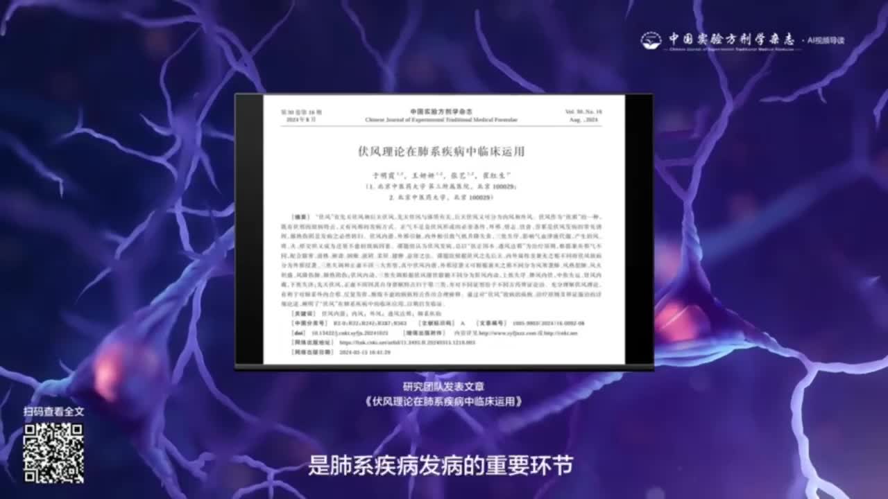


Different Effects of Fresh and Dried Dendrobium Huoshanense on Chronic Atrophic Gastritis
ObjectiveTo compare the protective effects of water extracts from fresh and dried


Editor-in-Chief:WU Yiling
Supervisor:
National Administration of Traditional Chinese Medicine
Sponsor:
Institute of Traditional Chinese Medicine,China Academy of Chinese Medical Sciences;China Association of Chinese Medicine
CN:11-3495/R
ISSN:1005-9903(Print)
ISSN:2097-1494(Online)
IF:5.474(CNKI)
Publication Cycle:Semimonthly
Address:No.16,Nanxiao Street,Dongzhimen,Dongcheng District,Beijing
Tel:010-84076882
E-mail:syfjx_2010@188.com
Hot word
 更多
更多
ObjectiveTo compare the protective effects of water extracts from fresh and dried


20 2024

18 Feb 2024

12 Jan 2024


 京公网安备11010802024621
